Stopping the amphibian apocalypse

Urgent Funding Appeal: Please help us to keep our amphibian project afloat.  

If you would like to discuss making a gift to support our work please email Brian Gratwicke gratwickeb[AT]si.edu . To make a credit card donation by phone please call our advancement department 202-633-8756.
Please mail checks to the address below and write Amphibian Rescue Project in the “Memo” field.

Attn: Amphibian Rescue Project
Smithsonian National Zoo and Conservation Biology Institute
P.O. Box 418320
Boston, MA 02241-8320

Check out this polka-dot color form of Vicente’s dart frog

Around 2004, a poison dart frog resembling the polka dot poison dart frog Oophaga arborea was smuggled into the European pet trade. The striking animal had yellow dots characteristic of the species and caused a sensation. Part of the excitement is because Oophaga arborea is a critically endangered species is from a very small known distribution where it inhabits bromeliads in the tree canopies. It was thought to be common in the 1980’s but may have declined due to chytridiomycosis. According to the IUCN redlist it was last seen in 2012, but was possibly heard from the Fortuna forest reserve more recently. Could this animal indicate that the polka dot poison dart frog survived the amphibian chytrid epidemic?

Small brown poison dart frog covered with with pale yellow dots

Male Oophaga vicentei photographed by Ariel Rodríguez

In June 2022, a population of frogs with yellow spots resembling the polka dot poison frog was discovered in Veraguas, outside the known distribution of the polka dot frog. The researchers collected a few specimens and analyzed them genetically where they most closely matched Vicente’s poison frog Oophaga vicentei. These dart frogs are known to be highly polymorphic, coming in slate gray, metallic blue, yellow or brick red with mottling. The yellow polka dot version, however, has now been confirmed to be Vicente’s poison dart frog as opposed to strawberry poison dart frogs Oophaga pumilio, or Oophaga arborea. Vicente’s poison dart frogs are listed by the IUCN as an endangered species, they can be locally abundant in places they are found and are known from a small area in Panama.

The paper was published in the journal Salamandra: Monteiro JPC, Ibáñez, R,  Mantzana-Oikonomaki, V.,  Pröhl, H.,  Rodríguez, A. (2023)  Genetic diversity of Oophaga vicentei (Anura: Dendrobatidae) and taxonomic position of a remarkable color morph from Panama. Salamandra 59 (4): 347-351

Amphibians remain the most imperiled vertebrate class and they need our help

Habitat destruction and disease are both well-documented causes of the decline of amphibians—among the most threatened animals on the planet—but a new paper analyzing two decades’ worth of data from around the world has found that climate change is emerging as one of the biggest threats to frogs, salamanders, and caecilians. The study was published today, Oct. 4, in the scientific journal Nature.

The study, “Ongoing declines for the world’s amphibians in the face of emerging threats,” is based on the second global amphibian assessment, coordinated by the Amphibian Red List Authority, which is a branch of the International Union for Conservation of Nature’s Species Survival Commission’s Amphibian Specialist Group, hosted and managed by Re:wild.

The assessment evaluated the extinction risk of more than 8,000 amphibian species from all over the world, including 2,286 species evaluated for the first time. More than 1,000 experts across the globe contributed their data and expertise, which found that two out of every five amphibians are threatened with extinction. These data will be published on the IUCN Red List of Threatened Species™.

Between 2004 and 2022, a few critical threats have pushed more than 300 amphibians closer to extinction, according to the study. Climate change was the primary threat for 39% of these species. This number is expected to rise as better data and projections on species’ responses to climate change become available. Climate change is especially concerning for amphibians in large part because they are particularly sensitive to changes in their environment.

Small red poison dartfrog called Andinobates geminisae carrying a tadpole on his back

Geminis’ dart frog is a critically endangered Panamanian amphibian that was only recently described. It is known from an area just 40km 2, males care for the offspring and transport tadpoles on their backs.

“As humans drive changes in the climate and to habitats, amphibians are becoming climate captives, unable to move very far to escape the climate change-induced increase in frequency and intensity of extreme heat, wildfires, drought and hurricanes,” said Jennifer Luedtke Swandby, Re:wild manager of species partnerships, Red List Authority coordinator of the IUCN SSC Amphibian Specialist Group, and one of the lead authors of the study. “Our study shows that we cannot continue to underestimate this threat. Protecting and restoring forests is critical not only to safeguarding biodiversity, but also to tackling climate change.”

Habitat destruction and degradation as the result of agriculture (including crops, livestock like cattle and livestock grazing, and silviculture), infrastructure development and other industries is still the most common threat, according to the paper. Habitat destruction and degradation affect 93% of all threatened amphibian species. Expanded habitat and corridor protection in the places most important for biodiversity is going to continue to be critical.

Disease caused by the chytrid fungus–which has decimated amphibian species in Latin America, Australia and the United States–and overexploitation also continue to cause amphibian declines. Habitat destruction and degradation, disease, and overexploitation are all threats that are exacerbated by the effects of climate change.

The study also found that three out of every five salamander species are threatened with extinction primarily as the result of habitat destruction and climate change, making salamanders the world’s most threatened group of amphibians. North America is home to the most biodiverse community of salamanders in the world, including a group of lungless salamanders abundant in the Appalachian Mountains of the eastern United States. Because of this, conservationists are concerned about a deadly salamander fungus that has been found in Asia and Europe, called Batrachochytrium salamandrivorans (Bsal), entering the Americas.

“Bsal has not yet been detected in the United States, but because humans and other animals can introduce the fungus to new places, it may only be a matter of time before we see the second global amphibian disease pandemic,” said Dede Olson, a research ecologist with the USDA Forest Service, member of the IUCN SSC Amphibian Specialist Group, and co-author on the paper. “It is critical that we continue to implement proactive conservation actions to prevent the spread of Bsal into the United States, including effective biosecurity practices for wild and captive amphibians, and rapid detection and response measures. The North American Bsal Task Force includes a multi-pronged strategic plan that includes: a continental surveillance and monitoring network; research studies identifying high-risk geographies and species; and collaborative partnerships across public, private, and governmental sectors.”

The Nature paper provides an update to the 2004 landmark paper that was based on the first global amphibian assessment for the IUCN Red List, which revealed the unfolding amphibian crisis for the first time and established a baseline for monitoring trends and measuring conservation impact. According to this new study, nearly 41% of all amphibian species that have been assessed are currently globally threatened, considered critically endangered, endangered or vulnerable. This is compared to 26.5% of mammals, 21.4% of reptiles and 12.9% of birds.

Four amphibian species were documented as having gone extinct since 2004—the Chiriquí harlequin toad (Atelopus chiriquiensis) from Costa Rica, the sharp snouted day frog (Taudactylus acutirostris) from Australia, Craugastor myllomyllon and the Jalpa false brook salamander (Pseudoeurycea exspectata), both from Guatemala. Twenty-seven additional critically endangered species are now considered possibly extinct, bringing the total to more than 160 critically endangered amphibians that are considered possibly extinct. The assessment also found that 120 species improved their Red List status since 1980. Of the 63 species that improved as the direct result of conservation action, most improved due to habitat protection and management.

“The history of amphibian conservation itself proves how vital this information is,” said Adam Sweidan, chair and co-founder of Synchronicity Earth. “If the IUCN Red List had been updated on a similar scale in the 1970s that it is today, we could have traced the sweeping amphibian disease pandemic 20 years before it devastated amphibian populations. It isn’t too late–we have this wealth of information, we have the Amphibian Conservation Action Plan, but plans and information are not enough. We need to act. We need to act fast.”

Conservationists will use the information from this study to help inform a global conservation action plan, to prioritize conservation actions at the global level, to seek additional resources, and to influence policy that can help reverse the negative trend for amphibians.

Citation: J.A. Luedtke, J. Chanson, K. Neam, L. Hobin et. al. 2023. Ongoing declines for the world’s amphibians in the face of emerging threats. Nature. https://doi.org/10.1038/s41586-023-06578-4 

Following reintroduced frogs

The Panama Amphibian Rescue and Conservation Project completed the first reintroduction trial of the Limosa Harlequin frog (Atelopus limosus) in 2017, and our findings were published in the journal Frontiers in Amphibian and Reptile Science. Reintroducing a species comes with a lot of unknowns and questions, just a few are: Where will the frogs go? What is their life like outside of a terrarium? Will they become infected with amphibian chytrid fungus? The purpose of this first reintroduction trial was to begin to unravel some of these questions so the researchers could adapt their strategies and improve the odds of the frogs in the wild.

We were able to get a detailed life of the frogs post-reintroduction by radiotracking the frogs and checking in on them daily. We found that when frogs were provided a 30-day acclimation period in a predator-free rainforest mesocosm, their probability of survival significantly increased and they did not disperse as far as the hard-released animals. We know from other studies that more movement can increase the likelihood of predators finding animals, and that likely happened in this study too. Frogs that were released without radio transmitters were 44x less likely to be reencountered during stream surveys (finding a frog in the rainforest isn’t easy!). We were able to follow the lives of these frogs in the wild for up to 56 days after release and developed a method that can use both radio-tracked animals and non-radiotracked animal encounters to estimate survivorship by assigning different detection probabilities in the model.

We observed a couple predation events of reintroduced frogs and some became infected with amphibian chytrid fungus. However, we learned a lot and has no shortage of questions to continue researching to get this species (and others in Panama) back into the wild.

Read the open access paper here:
Klocke, B., Estrada, A., Mataya, M., Medina, D., Baitchman, E., Belden, L., Guerrel, J., Evans, M., Baughman, J. and Connette, G., Illueca, E., Ibáñez, R., Gratwicke, B. (2023) Movement and survival of captive-bred Limosa Harlequin frog (Atelopus limosus) released into the wild. Frontiers in Amphibian and Reptile Science, 1, p.1205938. https://doi.org/10.3389/famrs.2023.1205938 

by Blake Klocke

First release trial of Atelopus limosus shows that animals rapidly recover a wild-type skin microbiome.

In 2017, Virginia Tech PhD students Angie Estrada and Daniel Medina conducted the first release trial of captive-bred Limosa Harlequin frogs at the Mamoni Valley Preserve. Their study aimed to closely observe how 1-year-old captive-bred animals would transition from captive conditions back into the wild. To soften the blow of the changing conditions, and to allow the researchers to capture the frogs again, they designed 30 square shaped mesocosms from plastic mesh. In reintroduction biology placing animals in a temporary field enclosure prior to release is known as a soft release. They filled the bottom layer with leaf litter collected from the forest floor that was rich in leaf-litter invertebrates, a diet quite different from the crickets and fruit-flies they are usually fed by staff at the Panama Amphibian Rescue and Conservation Center. Each mesocosm housed a single animal that was monitored daily  for a month and weighed and swabbed weekly.

It is known that captivity can alter skin microbiomes of amphibians and other captive animals, and that this is an important component of amphibian immune defenses. This study found that wild and captive Atelopus limosus had very different microbiomes, but that after a month living in mesocosms, the skin microbiome had rapidly changed to resemble the microbiome of wild individuals. One concern about bringing animals into captivity for prolonged periods is that animals might lose symbiotic microbes that help them to survive in the wild, which might reduce the fitness of captive-bred animals, but this study found that the skin microbiome was rapidly rewilded.

The mesocosms were a useful tool that protected the animals from larger predators, though one was killed by army ants. Female animals lost body condition more rapidly than males, but at the end of the trial their condition resembled that of wild-caught animals. About 15% of the animals became infected with the amphibian chytrid fungus in the first month compared to 13-27% Bd prevalence in wild amphibian community.

Estrada, A., Medina, D; Gratwicke, B, Ibáñez, R, Belden, L (2022) Body condition, skin bacterial communities and disease status: Insights from the first release trial of the Limosa harlequin frog, Atelopus limosus. Proceedings of the Royal Society B. https://doi.org/10.1098/rspb.2022.0586

New study genetically engineers skin bacteria in attempt to control the amphibian chytrid fungus

In our latest research project the Smithsonian Conservation Biology Institute and the Synthetic Biology Center Dept of Biological Engineering at MIT collaborated to test two technically challenging ideas using probiotic approaches to protect highly susceptible amphibians from the amphibian chytrid fungus (Bd). The paper was recently published in the Journal ISME Communications. We isolated a core frog skin bacteria that is found in high numbers on most golden frogs and genetically engineered it to produce an antifungal metabolite that kills the pathogen called violacein. By using a bacterium well-adapted to thrive on the frogs’ skin, that also produces antifungal metabolites we hoped to protect the frogs from disease. We were able to genetically modify one core skin microbe to produce violacein, but it did not persist well on the frog skin and was displaced after 4 weeks by the unmodified native bacteria strain. Treating the frogs with this genetically modified core skin microbe did not prevent the frogs from Bd or reduce infections.

In this new experiment, we tested two new probiotic strategies to protect frogs from Bd. 1) Using a consortium of antifungal bacteria isolated from the frogs 2) Using a core skin microbe found on all golden frog skin that was genetically modified to produce antifungal metabolites.

In a second experimental group we mixed a consortium of seven antifungal bacteria that had been isolated from golden frog skin and supplemented the skin microbiome with these potentially beneficial microbes. Three of the seven bacteria persisted on the skin after 4 weeks, but this probiotic treatment also failed to protect the frogs from disease. While these results are disappointing, we were able to successfully test two technically-challenging ideas that have been discussed in the amphibian conservation community for many years. Furthermore, this research illustrates some of the challenges we still face in understanding and manipulating microbiomes and in using synthetic biology to solve real environmental problems.

The research was led by Dr. Matthew Becker, Rob Fleischer and Brian Gratwicke (Smithsonian’s National Zoo and Conservation Biology Institute) and Dr. Jennifer Brophy and Christopher Voigt (Synthetic Biology Center Dept of Biological Engineering at MIT). Other collaborators include Ed Bronikowski, Matthew Evans, Blake Klocke, Elliot Lassiter, Alyssa W. Kaganer, Carly R. Muletz-Wolz (Smithsonian’s National Zoo and Conservation Biology Institute). Kevin Barrett (Maryland Zoo in Baltimore), Emerson Glassey & Adam J. Meyer (MIT). The work was funded by individual donors, the Smithsonian Institution Competitive Grants Program for Science, the Smithsonian Postdoctoral Fellowship, the U.S. Fish and Wildlife Service Division of International Conservation Amphibians in Decline Fund, the U.S. Defense Advanced Research Projects Agency’s Biological Robustness in Complex Settings program. The Maryland Zoo in Baltimore and the AZA Golden Frog Species Survival Program provided surplus-bred animals for research.

By Brian Gratwicke

Becker, M.H., Brophy, J.A.N., Barrett, K, Bronikowski, E., Evans, M., Glassey, E., Klocke, B. Lassiter, E., Meyer, A.J., Kaganer, A.W., Muletz-Wolz, C.R.,  Fleischer, R.C., Voigt, C.A., and Gratwicke, B. Genetically modifying skin microbe to produce violacein and augmenting microbiome did not defend Panamanian golden frogs from disease. ISME Communications


Meet the spectacular Borderlander frog Atelopus fronterizo, Panama’s newest frog species!

Milan Vesely and Abel Batista scientifically described Panama’s seventh harlequin frog species in the journal Zoological Research in April 2021. This beautiful harlequin frog species has been in biological collections for some time, the first museum specimen was first collected by Henri Pittier at Puerto Obaldlia in 1911, but has previously been assumed to be related to other sister species that it closely resembles. Historically collected formalin-preserved specimens were unsuitable for genetic analyses and so the frog remained undescribed.

Vesely and Batista conducted expeditions to the Darien to collect new specimens and this allowed them to conduct genetic analysis showing that this species is most closely related to Atelopus certus and Atelopus glyphus, but is genetically distinct enough to warrant recognition as a species. It also has a slightly different call and morphological characteristics that are also described in the paper.

Borderlander Harlequin Frog

Atelopus fronterizo Photo by Abel Batista, UNACHI-Fundación Los Naturalistas-SNI (SENACYT)

They named the frog Atelopus fronterizo to refer to borderland inhabitants and the Panamanian border security force who protect the Darien mountain range in NE Panama on the border where this frog is found. Like other harlequin frog species in Panama, even though they live in well-protected habitat, they are Likely Critically Endangered due to the threat of the amphibian chytrid fungus that has caused the declines of other Atelopus species in Panama.

Vesely, M. and Batista, A., 2021. A new species of Atelopus (Amphibia: Bufonidae) from eastern Panama. Zoological research, pp.272-279.

Spindly leg syndrome is reduced by increasing calcium hardness of water used to rear tadpoles

Atelopus varius metamorph with spindly leg (left) and without spindly leg (right)

Rearing frogs in captivity has its own unique challenges, one problem that has been a persistent issue in the Panama Amphibian Rescue and Conservation Project is spindly leg syndrome (SLS). This common musculoskeletal disease is mostly associated with captive amphibian breeding. SLS is a condition where legs of newly metamorphed amphibians, with otherwise healthy and typical development, are poorly developed and cannot support the weight or newly metamorphed froglets. Ultimately, SLS leads to death as the animal is unable to move or feed themselves. A brief review online will reveal a host of theories and potential remedies for the condition ranging from parental nutrition to water quality and dietary supplements, but there are very few replicated peer-reviewed experiments identifying the cause of this disease.

Elliott Lassiter and Orlando Garcés with the experimental rearing setup

As an intern with the Panama Amphibian Rescue and Conservation Project I teamed up with Orlando Garcés a graduate of the University of Panama and employee of the project to conduct an experiment primarily funded by the Morris Animal Foundation. We had observed that SLS was most prevalent in water that did not have any supplementary calcium and we knew that incoming water to our facility was very soft (lacking in calcium hardness). Bone growth is the symptom of SLS, therefore, we decided to look at the principle minerals affecting bone growth: calcium and phosphate. Tadpoles can gain calcium through their diet but they absorb about 70% of their calcium from the water through their gills and skin. The collected calcium is then stored in endolymphatic sacs in their heads and used during metamorphosis when tadpoles’ skeleton turns from cartilage into bone and limbs begin to grow.

We took 600 Atelopus varius tadpoles and divided them into three calcium treatments (low, medium, high) and then divided those into two groups one with added phosphate and one without added phosphate.  We monitored our tadpoles until they metamorphosed, at which point we looked at their legs and body posture to determine whether or not they had SLS. We found that calcium supplementation drastically increased survivorship overall and that the medium and high calcium groups had less SLS than the low calcium groups. Addition of phosphate also decreased the prevalence of SLS in low calcium treatment.

Based on the results of this study we were able to determine that SLS in harlequin frogs, is linked to an imbalance in calcium and phosphate homeostasis. Therefore, our current husbandry recommendation to reduce SLS in frogs and toads is to consider checking water hardness to determine if it is too soft. We also advise against over feeding tadpoles which has been shown to cause an increase in SLS prevalence in another experiment. We hope that our findings can guide future SLS research and help to lower the prevalence of SLS in captive amphibians, improving animal welfare. This research will help to improve the long-term sustainability of captive populations while researching solutions for the amphibian chytrid fungus and eventual reintroduction of these frogs back into the wild.

Lassiter, E., Garcés, O., Higgins, K., Baitchman, E., Evans, M., Guerrel, J., Klaphake, E., Snellgrove, D., Ibáñez, R. and Gratwicke, B., 2020. Spindly leg syndrome in Atelopus varius is linked to environmental calcium and phosphate availabilityPloS one15(6), p.e0235285.

By Elliot Lassiter and Orlando Garcés

A vigorous immune response to the chytrid fungus is associated with susceptibility to the disease

For frogs dying of the invasive chytridiomycosis disease, the leading cause of amphibian deaths worldwide, the genes responsible for protecting them may actually be leading to their demise, according to a new study published today in the journal Molecular Ecology by Smithsonian Conservation Biology Institute (SCBI) and University of Central Florida researchers.

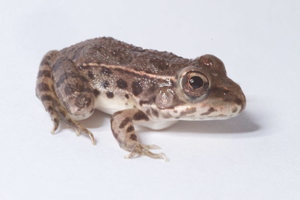
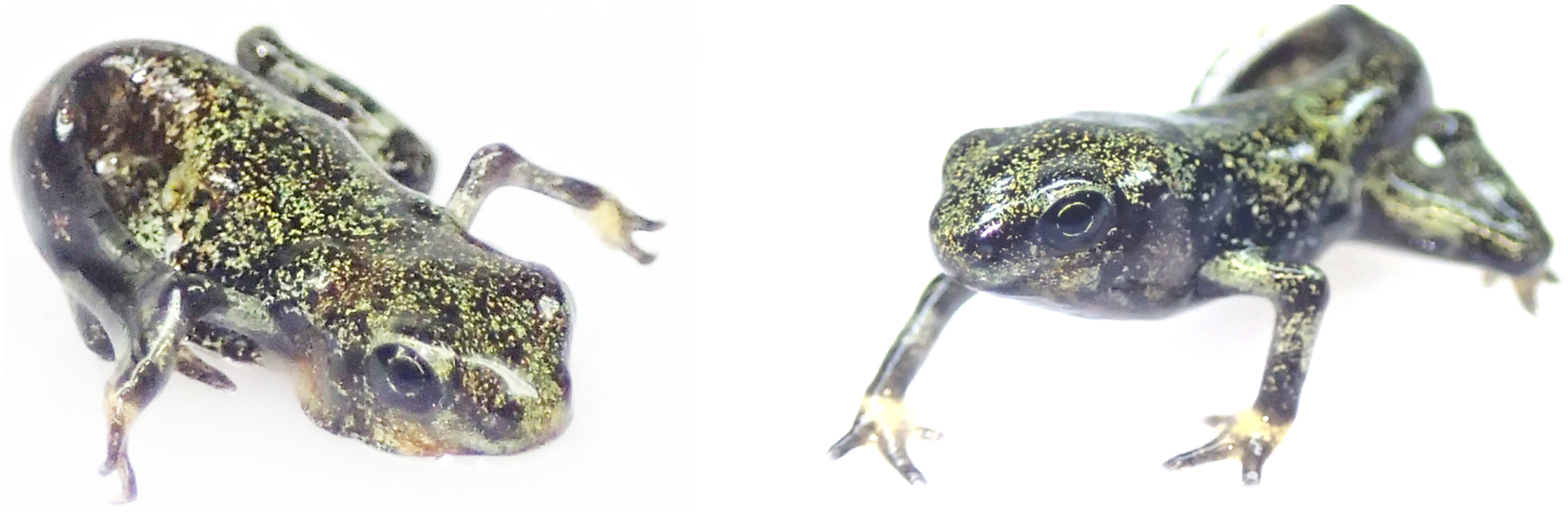
The lowland leopard frog, found in river drainages in Arizona, is one of a few amphibian species in which some individuals survive infection by Batrachochytrium dendrobatidis chytrid fungus (Bd) while other individuals do not—even when they live in the same local population.

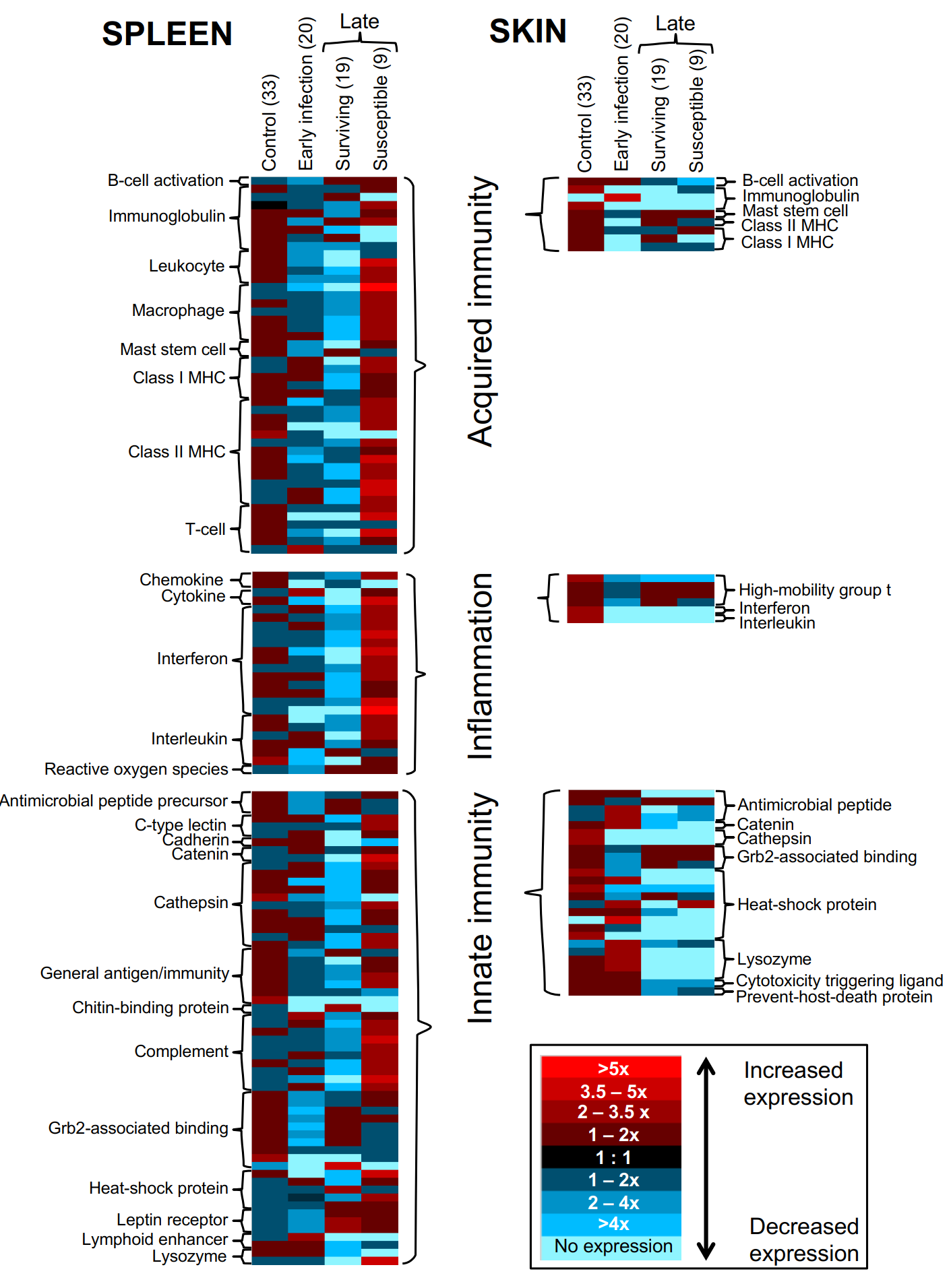
In a study of lowland leopard frogs infected with Bd, the fungus that causes the disease chytridiomycosis or chytrid, researchers found that frogs that died from the disease had higher expression of major histocompatibility complex and other immune system genes than frogs that survived it. Those genes help organisms fight off infections and foreign substances.

“This result was totally counterintuitive and the opposite of the pattern we expected to recover,” said Anna Savage, the study’s lead author, an associate professor in UCF’s Department of Biology and former postdoctoral fellow at SCBI’s Center for Conservation Genomics (CCG).

Comparison of differential gene expression in control, early infected, surviving and susceptible frogs in both the spleen (left) and skin (right). 

“My previous research on these immune genes showed that some variants were associated with higher survival to Batrachochytrium dendrobatidis, so I hypothesized that those genes were enabling the frogs to have a stronger immune response that would kill the fungus,” she said. “Instead, it seems like those stronger responses are linked to susceptibility, and the genes associating with survival are linked to reduced immune function.”

Savage said acquired immune responses can be very potent, require a lot of energy from the body and can sometimes produce toxic byproducts that harm the host and the pathogen.

“Immune responses are much more complex than just an on-off switch,” she said. “A big part of the immune system is regulating the type, timing and dosage of a particular response, and if any of those components get dysregulated, it can have extremely negative consequences.”

She said, for instance, Batrachochytrium dendrobatidis suppresses the host immune system by killing B and T lymphocytes. “Because those are the same cells that proliferate during acquired immune responses, producing lots of those cells might just be wasting energy on something that chytrid can easily destroy,” she said.

Amphibian populations are in decline around the world, with two-thirds of the world’s 8,000 species considered to be threatened and nearly 200 species that have already gone extinct in the last two decades. In the U.S., amphibian populations overall are declining at a rate of nearly 4 percent a year, with some areas, such as the Rocky Mountains and the West Coast, facing a higher rate of decline, according to the U.S. Geological Survey.

Although the researchers studied immune gene expression in lowland leopard frogs with chytridiomycosis, the findings may be useful for studying the disease in other frog species due to genetic similarities they share, Savage said.


Lowland leopard frogs were chosen for the study because their responses to chytridiomycosis vary from one individual to the next, unlike many other frog species that are completely susceptible to the disease or are completely resistant or tolerant.

This allowed the researchers to rule out genetic variation between species and pinpoint specific differences in lowland leopard frogs’ immune genes that predicted different responses to infection.

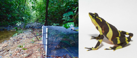
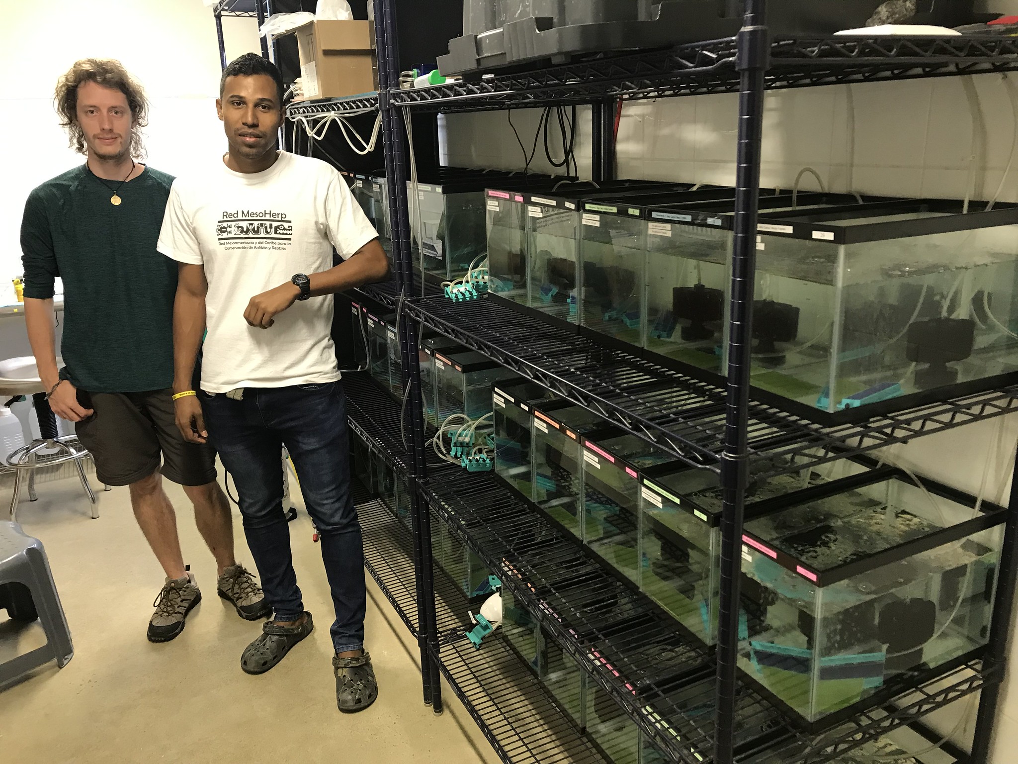
The frogs were collected in Arizona and shipped overnight to the Smithsonian’s National Zoo in Washington, D.C., where the infection experiments were conducted. Subsequent analyses of gene expression occurred at the SCBI’s Center for Conservation Genomics. Statistical analyses of the data were performed at UCF.

Robert Fleischer, senior scientist and head of the SCBI’s CCG, co-authored the study and was Savage’s main advisor for the research when she was a postdoctoral fellow at the Smithsonian. Fleischer said the results help in understanding why some frogs survive the disease and others do not.

“If we can solve this mystery, and we have taken a big step in that direction with this study, our hope and plan is to use this information to develop resources and strategies to mitigate the disease in the more susceptible species, and to counter the worldwide tide of extinction and endangerment caused by chytrid,” he said.

The researcher said the findings also show that acquired immune responses, such as those generated by vaccination, may not always be useful in combating invasive diseases of conservation concern.

Brian Gratwicke, a conservation biologist with SCBI; Katherine Hope, an associate veterinarian with the Smithsonian’s National Zoo; and Ed Bronikowski, senior curator of the Smithsonian’s National Zoo, were study co-authors as well.

The research was funded by a Smithsonian Institution Competitive Grants Program for Science grant, the Smithsonian’s Center for Conservation Genomics and a Smithsonian Institution Molecular Evolution Postdoctoral Fellowship.

Anna E. Savage, Brian Gratwicke, Katharine Hope, Edward Bronikowski, Robert C. Fleischer. Sustained immune activation is associated with susceptibility to the amphibian chytrid fungusMolecular Ecology, 2020; DOI: 10.1111/mec.15533

written by Robert Wells (University of Central Florida)